
Magnetic sensor is basis for image diagnosis technology
Yokohama National University, Japan and TDK have developed a prototype image diagnosis technology using a high-sensitivity magnetic sensor.
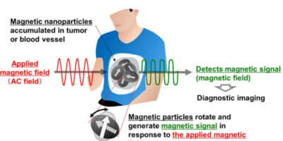
The prototype technology is related to the magnetic particle imaging method which is intended to detect and create images of magnetic particles accumulated in a tumour or blood vessel.
Magnetic resonance imaging (MRI) diagnostics and X-ray computerised tomography (CT) scanning are used in clinical services in the diagnosis of organ health, tumours and other conditions using the contrasting density of imaged objects. In contrast, the magnetic particle imaging is intended for use in detecting only the tracers in the imaged objects to create images similar to positron-emission tomography (PET) and similar technologies.
Magnetic particle imaging is used to detect the magnetic signals generated by magnetic particles accumulated in a tumour or blood vessel from outside of the body. In medical imaging, it is important for devices to be extremely sensitive to detect small amounts of magnetic particles. Magnetic particle imaging technologies primarily use a method that measures electromotive force electromagnetically induced through detection coils but the new technology developed by the university uses a prototype high-sensitivity magnetic sensor developed by TDK for use in the detection weak magnetic fields and at room temperature.
The prototype sensor has been shown to measure magnetic field distribution in a heart. Through this development, the sensor has reduced the strength of the alternate current (AC) magnetic fields applied from outside of the body to one 10th lower than conventional levels, reported TDK and Yokohama National University. The reduced strength of the applied field is achieved by the non-linear response characteristics of the sensor to the measured magnetic field strength.
Based on this, it is expected that the utilisation of high-sensitivity magnetic sensors will enable magnetic particles to be detected across wider imaging ranges including the head or whole body of a human.
Yokohama National University and TDK will continue to develop this technology, with the goal of creating magnetic particle imaging devices that can be used practically in clinical services.
Yokohama National University (YNU) has brought together faculty members with diverse specialties such as humanities, social sciences, science, and engineering at the university’s One Campus to provide education and research. It aims to become a world-class research university, collaborating with various industries, regions, communities, and citizens, both domestically and internationally, across disciplines.
TDK was established in 1935 to commercialise ferrite, a key material in electronic and magnetic products. TDK’s portfolio features passive components such as ceramic, aluminium electrolytic and film capacitors, as well as magnetics, high-frequency, and piezo and protection devices. It also includes sensors and sensor systems such as temperature and pressure, magnetic, and MEMS sensors. TDK also provides power supplies and energy devices, marketed under the product brands TDK, Epcos, InvenSense, Micronas, Tronics and TDK-Lambda. TDK focuses on demanding markets in automotive, industrial and consumer electronics, and information and communication technology. The company has a network of design and manufacturing locations and sales offices in Asia, Europe, and in North and South America.